Jul 13, 2026 | Blog Post
The clinical challenge
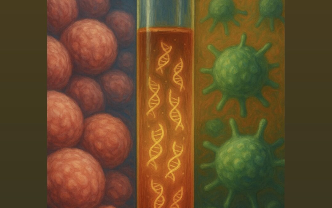
Liquid biopsies have undoubtedly reshaped precision oncology. A simple blood draw can now reveal the molecular blueprint of a patient’s cancer, and this information can be subsequently used to guide treatment decisions. Actionable mutations, resistance mechanisms, and tumor evolution can all be evaluated without the need for an invasive tissue biopsy. But as the field has matured, so has our appreciation for a persistent confounder lurking in the very same blood sample we draw: clonal hematopoiesis.
As we age, hematopoietic cells accumulate somatic mutations, some of which are in cancer-related genes and can lead to increased cell proliferation. This phenomenon is called clonal hematopoiesis (CH) and is remarkably common. By some estimates, over 40% of individuals with advanced cancers harbor at least one CH variant among clinically reportable genes, and the prevalence increases steeply with age.
The problem? Many of these CH-derived mutations sit in the very same genes we use to match patients to targeted therapies: BRCA1/2, ATM, TP53, and others. To put this in clinical context, when a variant is detected in a patient’s blood, the clinician must determine whether it is a bona fide tumor alteration that warrants targeted therapy or a confounder arising from a hematopoietic clone unrelated to the cancer itself. Misinterpreting this could result in administering the incorrect medication.
Ways to alleviate these challenges include matched tumor or white blood cell (WBC) sequencing. If the variant is present in both circulating free DNA (cfDNA) in plasma and WBC DNA, it provides strong evidence that the variant is CH-derived. However, in routine clinical practice, tumor or WBC sequencing adds cost, complexity, and turnaround time. Consequently, physicians may need to make treatment decisions based on detected cfDNA variants, without complete confidence that those variants are originating from the cancer they are trying to treat.
This issue is tackled using a new machine learning model named plasmaCHORD.
What is plasmaCHORD?
plasmaCHORD (plasma Clonal Hematopoiesis ORigin Detection) is a machine learning model designed to determine, for each variant detected by a cfDNA targeted fixed-gene panel sequencing, whether it originated from tumor cells or from WBCs. PlasmaCHORD combines information across three levels:
1. Mutant cell-free DNA fragment-level features: structural characteristics of the cfDNA fragments carrying the variant, leveraging the differences in physical properties between tumor-derived and hematopoietic-derived cfDNA.
2. Variant-level features: properties of the mutation itself, including its genomic context and known prevalence in CH versus solid tumors.
3. Gene-level features: evaluation of whether the gene carrying the mutation is frequently mutated in CH versus solid tumors.
3. Patient-level features: clinical characteristics that may influence the probability of CH, such as age of the patient.
By combining these inputs, plasmaCHORD captures the complexity of cfDNA biology in the context of a specific host in a way current rule-based filters cannot.
How we built and validated it
We developed plasmaCHORD using a training set of 426 variants identified by next-generation sequencing of cfDNA from 225 patients with stage I–IV solid tumors. Critically, the origin of each variant, tumor, or CH was determined by matched sequencing of WBCs and tumor tissue. After fine-tuning the model parameters, we tested plasmaCHORD on an independent validation cohort of 1,418 plasma variants from 114 patients with metastatic cancers. The model maintained strong performance, with an area under the curve (AUC) of 0.94 in the training set and a similar AUC of 0.90 in the validation set. Importantly, plasmaCHORD demonstrated a significant improvement in accuracy, particularly for clinically significant genes that matter most for treatment decisions.
From computation to bedside
But what might the clinical impact of plasmaCHORD look like? We applied plasmaCHORD to cfDNA sequencing data from patients enrolled in a prospective precision oncology clinical trial (NCT05585684). We highlighted two challenging cases in which actionable variants were detected but the origin was ambiguous, raising the possibility that targeted therapy could be inappropriate or ineffective. PlasmaCHORD correctly classified these variants as tumor- or CH-derived, helping to avoid inappropriate genotype-based treatment decisions and to accurately identify patients who might otherwise have been incorrectly matched to a targeted therapy.
The bigger picture
The challenge of CH in liquid biopsies is not going away; if anything, it is growing. As liquid biopsy panels expand to cover more genes, as comprehensive genomic profiling moves into earlier disease stages, and as the population of cancer patients ages, the risk of CH-driven misinterpretation will only increase. Approaches like plasmaCHORD can bridge that gap, adding a layer of intelligent interpretation to the thousands of plasma-only cfDNA sequencing assays conducted each year. PlasmaCHORD is therefore a computational safeguard, ensuring that when we act on a variant, we are acting on the right signal.
Read the full paper here.
Jul 9, 2026 | Blog Post
Precision oncology is increasingly driven by next-generation sequencing (NGS) technologies, advanced techniques that allow for parallel sequencing of millions of nucleic acid fragments. These technologies have enabled comprehensive genomic profiling of tumor tissue and circulating cell-free DNA (cfDNA), the latter facilitating minimally invasive molecular testing in individuals with cancer. cfDNA consists of short DNA fragments released into the bloodstream; a fraction of which originates from dying cancer cells and is referred to as circulating tumor DNA (ctDNA). Despite the promise of liquid biopsies, interpreting their results can be challenging because not all DNA molecules detected in the bloodstream originate from a tumor. One important confounding factor in liquid biopsies is clonal hematopoiesis (CH), an age-related process in which blood cells acquire mutations and expand over time. These blood-derived mutant DNA molecules are released into the bloodstream alongside tumor-derived DNA, making it difficult to determine whether a mutant DNA molecule originated in cancer or non-cancerous blood cells. Given that precision oncology increasingly relies on genomic biomarkers to guide treatment selection and clinical trial enrollment, the accurate classification of mutations detected in liquid biopsies is critical for therapeutic decision-making.
To address these challenges and enhance the accuracy of liquid biopsies, recent work from our Molecular Oncology laboratory introduced plasmaCHORD, a machine learning model that accurately distinguishes tumor-derived mutations from CH mutations in cfDNA. This work was presented at the 2026 American Association for Cancer Research Annual Meeting, was recognized with the AACR-Margaret Foti Foundation Scholar-in-Training Award and was concurrently published in Clinical Cancer Research.
Several approaches can help resolve the true origin of mutations detected in cfDNA. Matched white blood cell (WBC) sequencing can identify variants present in blood cells, allowing these germline and CH-derived variants to be filtered from cfDNA results. Similarly, matched tumor next-generation sequencing can help confirm which plasma variants are truly tumor-derived. However, both approaches require additional sequencing and may be limited by cost, tissue availability, or the feasibility of obtaining tumor tissue in patients with metastatic cancer. plasmaCHORD was developed to address this gap by directly predicting variant origin from plasma NGS data alone.
To train plasmaCHORD, three types of information were integrated: features of the DNA fragments carrying each mutation, features of the mutation and gene itself, and patient-level characteristics. Fragment-level features included DNA fragment length and cleavage-site location, while mutation-level features included variant allele frequency and gene-level context. Patient age was also included because clonal hematopoiesis becomes more common with aging. Using these inputs, plasmaCHORD was trained to classify plasma cfDNA mutations as either tumor-derived or CH-derived. The model was initially trained in patients with both localized and metastatic cancers, and a set of 426 mutations with known origins. It was subsequently validated using an independent dataset of 1,418 mutations of both tumor and CH origin.
Overall, plasmaCHORD demonstrated strong performance in predicting whether plasma cfDNA mutations were tumor-derived or CH-derived. In the independent validation cohort, the model achieved an area under the curve (AUC) of 0.902 and approximately 81% accuracy, indicating that it could reliably distinguish between the two mutation origins. Importantly, plasmaCHORD performed consistently across a diverse range of cancer types, including those not represented in the training cohort. The study also showed that no single feature was sufficient to classify the origin of mutations on its own. Instead, performance depended on integrating multiple sources of information. The most informative features included a measure of how often a gene is mutated in blood-related cancers compared to solid tumors, DNA fragment length metrics, and patient age. plasmaCHORD also performed well in particularly challenging settings, including mutations in commonly mutated cancer driver genes including TP53, where it achieved accuracies of 82.8% and 76.9%, respectively.
To demonstrate plasmaCHORD’s utility in real clinical scenarios, the authors applied the model to two challenging cases reviewed by the Johns Hopkins Molecular Tumor Board. In one patient with metastatic ALK fusion-positive NSCLC, plasma-only liquid biopsy detected an ATM truncating mutation. If tumor-derived, this finding could have raised consideration of DNA repair–targeted therapy; however, the Molecular Tumor Board determined that the mutation was likely CH-derived. In a second patient with NSCLC, liquid biopsy identified an EZH2 truncating mutation that could have indicated potential eligibility for EZH2-directed therapy if tumor-derived. plasmaCHORD classified both variants as CH-derived, matching the Molecular Tumor Board’s interpretation and subsequent confirmation by matched white blood cell sequencing.
Our study highlights that CH-derived mutations are not limited to canonical CH-associated genes. Some suspected CH variants occurred in clinically relevant driver genes and known tumor hotspots, underscoring the limitations of simple gene-based filtering strategies in cfDNA mutation profiling. To this end, plasmaCHORD has the potential to improve the accuracy and clinical utility of plasma comprehensive genomic profiling without requiring matched white blood cell or tumor NGS. Refinement of the plasmaCHORD method and incorporation of additional ctDNA fragment features may further improve performance in a wide range of cancers. Importantly, the model offers an innovative, practical approach for routine liquid biopsy analysis in clinical care. As liquid biopsies become increasingly integrated into cancer care, methods that improve the identification of true tumor-derived mutations may enhance biomarker discovery and support more informed treatment decisions.
Sep 10, 2025 | Blog Post
Diffuse pleural mesothelioma remains a uniquely challenging malignancy. Its growth pattern undermines conventional imaging, its biology yields scant circulating tumor signal, and debate exists around the value and timing of surgery. Against this backdrop, we asked two simple questions of central practical importance and clinical significance. First, can immune checkpoint blockade be given before and after surgery without derailing the operative plan or compromising safety in patients with resectable pleural mesothelioma? Second, can an ultra-sensitive, tumor-informed liquid biopsy provide a clinically meaningful molecular read-out of peri-operative immunotherapy outcomes, early enough to guide therapeutic decision making?
Patients with good performance status and multidisciplinary confirmation of operability received either neoadjuvant nivolumab alone or combined with a single dose of ipilimumab. All patients who underwent resection also received post-operative therapy, including adjuvant nivolumab for one year per protocol. The co-primary endpoints were feasibility, defined a priori as proceeding to surgery without protocol-defined delay, and safety, captured by dose-limiting toxicities during the neoadjuvant window. Progression-free and overall survival were evaluated as exploratory outcomes alongside tumor-informed, whole-genome sequencing liquid biopsy, measuring circulating tumor DNA (ctDNA) at baseline, through neoadjuvant therapy, immediately pre-operatively, and again post-operatively.
The clinical verdict on feasibility and safety was clear. More than four-in-five patients in each arm reached the operating room as planned, and complete macroscopic resection was achieved in most cases. Immune-related toxicities aligned with expectations for immunotherapy agents, and adjuvant nivolumab was generally well-tolerated. These findings address the core, practical anxiety that neoadjuvant immunotherapy might derail operability, which it did not.
Efficacy signals, while exploratory in this phase 2 clinical trial setting, were encouraging and coherent. Median progression-free and overall survival favored the combination of nivolumab with ipilimumab over nivolumab alone, and a meaningful fraction of patients in the combination arm remained alive and recurrence-free at the time of data analysis. One should avoid over-interpreting arm-to-arm contrasts in this small, sequentially-accrued study, but the pattern – durable control in a subset without dramatic radiographic responses – foreshadows the central translational lesson of this work.
That lesson is that, in mesothelioma, cell-free tumor DNA detection in the bloodstream is a more reliable indicator of treatment effect than tumor measurements on imaging. Conventional response rates by modified RECIST were modest, as expected in this pleural surface-spreading cancer type, which often thins rather than shrinks; yet the ctDNA assay drew a much sharper picture. By leveraging genome-wide mutation profiles from each patient’s tumor, and utilizing a machine learning framework to suppresses technical noise in cell-free DNA, we were able to detect extraordinarily low tumor levels and track ctDNA dynamics over time. Two clinically intelligible patterns emerged.
First, detectable ctDNA during the neoadjuvant window correlated with the feasibility endpoint at the heart of this trial. Patients who ultimately could not complete surgery – either because their disease progressed between planning and incision, or because the tumor proved unresectable intraoperatively – showed persistent or rising ctDNA levels, while still being nominally eligible on imaging. This is the kind of actionable information that can promptly redirect specific patients to alternative treatment strategies.
Second, ctDNA served as an indicator of long-term progression-free survival. Patients with undetectable ctDNA by the end of neoadjuvant therapy and immediately before surgery experienced substantially longer progression-free survival than those with residual ctDNA; this same pattern held, often more strongly, when one looked at two-timepoint dynamics. Significant drops in tumor fraction from baseline, or persistently undetectable ctDNA throughout the neoadjuvant course, were observed in patients most likely to attain durable control after surgery; additionally, post-operative re-emergence of ctDNA predicted disease recurrence. Importantly, these molecular trajectories frequently diverged from scan-based impressions. In other words, ctDNA captured the direction and velocity of disease progression and therapy response when imaging techniques could not.
It is worth pausing on what, precisely, we can and cannot claim based on these data. The notion that ctDNA is prognostic, i.e., that it stratifies patients by risk independent of treatment choice – is strongly supported here, since those without detectable ctDNA at key junctures attained better outcomes. Whether ctDNA is also predictive, i.e., whether its clearance specifically marks benefit from neoadjuvant checkpoint blockade, is a more nuanced claim in a non-randomized phase 2 study without a non-immunotherapy control arm. The temporal coupling of ctDNA reduction to neoadjuvant dosing, and the association with improved clinical outcomes, together make a persuasive case that the ctDNA signal is treatment-linked. However, definitive proof will require larger, prospective trials, adequately powered to prove the clinical benefit of a ctDNA-guided adaptive therapy.
Two practical implications follow immediately. First, the trial establishes that neoadjuvant checkpoint blockade can be considered among curative-intent therapy options for resectable mesothelioma without jeopardizing surgery. Second, ctDNA residual disease after neoadjuvant immunotherapy and prior to surgery – offers a crisp, quantitative, molecular read-out that may refine individual care. Clear molecular response supports proceeding to resection, while a persistent ctDNA signal should trigger a re-consideration of medical management. After resection, reappearance of ctDNA should prompt intensified surveillance and the swift deployment of systemic therapy.
No early-phase study is without limitations. Arms were accrued sequentially, and the participating sites brought deep surgical expertise that not all centers can immediately replicate. Mesothelioma’s low mutational burden and locoregional spread will always challenge liquid biopsy analytical and clinical sensitivity. Yet even within these constraints, the core conclusions hold; perioperative immunotherapy is feasible and safe, and ctDNA provides an accurate and timely account of tumor burden dynamics and therapeutic response – something imaging alone has not reliably delivered in this setting.
Taken together, the purpose of this trial was to determine whether perioperative immune checkpoint blockade is practical in resectable mesothelioma, and whether an ultra-sensitive tumor-informed ctDNA assay can serve as an early, clinically-useful biomarker. The result is yes on both counts. The therapy was feasible and safe, and the liquid biopsies were not merely measurable, but clinically informative, identifying patients likely to attain clinical benefit, signaling those at risk of surgical attrition or early recurrence, and offering a path to more adaptive, individualized, and precise perioperative care. As the field moves to larger studies, these findings provide a clear blueprint; keep immunotherapy in the perioperative conversation, and let ctDNA, measured carefully and interpreted judiciously, guide the rhythm of that dialogue.
Dive deeper and read the full study here: https://www.nature.com/articles/s41591-025-03958-3

Aug 18, 2025 | Blog Post
Precision oncology has fundamentally reshaped the treatment paradigm within the field, with individualized biomarker-matched therapies enhancing the arsenal oncologists use to manage cancer.
For the evolving ecosystem of precision oncology to advance toward a nationwide integration, efforts should focus on expanding access to genomic expertise, molecular tumor boards, and clinical trial infrastructure – resources that remain scarce outside academic centers. There thus arises a need to shift the strategic approach to advancing precision oncology so that it emphasizes not only technological and pharmacological progress but also clinical utility and broader accessibility.
Significant efforts have been made toward establishing comprehensive molecular profiling in first-line decision-making, where clinically appropriate. Non–small cell lung cancer (NSCLC) has long exemplified the integration of baseline molecular testing; where genomic alterations such as EGFR mutations, ALK and ROS1 rearrangements, MET exon 14 skipping, BRAF V600E mutations, and PD-L1 expression levels are systematically assessed to guide personalized therapy. Similarly, in breast cancer, estrogen receptor (ER) and progesterone receptor (PR) expression, and epidermal growth factor receptor 2 (HER2) expression/amplification are predictive biomarkers evaluated at baseline. Furthermore, tumor genotyping for PIK3CA and ESR1 mutations has become clinically actionable, with FDA approvals of specific drug classes for these biomarker-defined subsets of hormone receptor-positive breast cancer. These models illustrate that when actionable biomarkers are implemented, testing informs therapeutic decision-making and improves clinical outcomes.
In terms of real-time monitoring of tumor burden and therapeutic response, liquid biopsy analyses have emerged as paradigm-shifting technologies; longitudinally assessing circulating tumor DNA (ctDNA) dynamics and evaluating residual disease across stages and therapies. Liquid biopsies also allow for tracking tumor evolution, detecting emerging resistance, elucidating mixed responses or stable disease, and identifying recurrence before it becomes radiographically or clinically apparent.
Establishing precision oncology as the standard of care, however, requires two key elements: technology to advance cancer discovery and education to scale its impact. By providing expert molecular interpretation and implementing decision-support tools in community clinics, we can expand access and enrolment in biomarker-matched therapies and clinical trials.
At this precise intersection – between education and technology – we are building a precision-oncology decision-support platform embedded within the Johns Hopkins Molecular Tumor Board to standardize evidence assessment and expand equitable access to expert interpretation and recommendations. In line with the scope of the NCI Community Oncology Research Program – which bridges clinical trials and care delivery research with community clinics – we envision that our initiatives will help operationalize precision oncology and create clear on-ramps to clinical trial screening and enrolment.
Outreach programs tailored for community oncologists, improved referral networks, and regionally or virtually hosted case-based molecular tumor boards (MTBs) have further democratized access to genomic expertise and improved clinician confidence in navigating molecular findings. Importantly, these educational models should be sensitive to workflow realities in high-burden settings and emphasize the clinical utility of precision oncology approaches. Expanding genomic literacy beyond the physician level to include nurse navigators, genetic counsellors, and case managers can further support patient-centered education, and increased engagement in shared decision-making processes, including clinical trial participation.
Internationally, promising models are emerging. Regional MTBs supported by international consortia have demonstrated feasibility and clinical benefit even in resource-constrained settings, providing a blueprint for scalable transnational and supranational precision oncology frameworks.
The future of oncology depends not only on identifying new biomarkers and developing advanced targeted therapies, but also on breaking down the educational and logistical barriers that restrict their access. Through the meaningful work of precision oncology groups, including our own at the Johns Hopkins MTB, we are hopeful that soon precision oncology will evolve from a specialized innovation into an accessible standard, embedded across all levels and stages of the cancer care continuum.
Jul 27, 2025 | Blog Post
Over the past decade, significant progress has been achieved in treating lung cancer, particularly for individuals with non-small cell lung cancer (NSCLC). In particular, the development of immune checkpoint inhibitors – which harness a patient’s immune system to fight cancer – has resulted in significant improvements in patient outcomes. Immune checkpoint inhibitors work by blocking inhibitory molecules that cancer cells use to evade immune surveillance, enabling the patient’s immune system to target cancer cells. This therapeutic approach is entirely unlike chemotherapy, which targets all rapidly dividing cells and thus has the potential to target some healthy cells – e.g., hair cells and cells in the lining of the digestive tract.
Still, not all patients attain clinical benefit from these novel immunotherapies. Some lung cancers harbor an “immunologically cold” phenotype, meaning they are more likely to evade immune recognition and elimination by the immune system even when treated with immune checkpoint inhibitors. For example, tumors with fewer somatic mutations, which are considered to have a low tumor mutation burden (TMB), and tumors with no PD-L1 expression, generally show a poorer response to immunotherapy. This raises the question of how we can improve responses to immunotherapy in patients with tumors that are predisposed to resistance.
Preclinical evidence suggests that radiation therapy (RT) could help circumvent immunotherapy resistance. When cancer cells are subjected to RT, they release antigens into the nearby environment, known as the tumor microenvironment (TME). The immune system can then recognize these antigens and tailor immune responses to the molecular “blueprint” of the cancer. In this way, RT may be able to enhance anti-tumor immune responses, both at the primary tumor treated with radiation and systemically, and at distant metastases far from the site of radiation. This effect is known as the abscopal effect of radiation (‘ab’ meaning away from, and ‘scopus’ meaning target).
These findings suggest that RT could enhance the response to immunotherapy, even at distant metastatic sites. However, this effect has not been consistently shown, especially in human biospecimens. To bridge this gap, we studied how lung cancers responded to immunotherapy in a phase 2 randomized clinical trial of patients with NSCLC receiving combination radioimmunotherapy. In one arm of this trial, patients received radiotherapy together with pembrolizumab, while in the control arm, patients received pembrolizumab alone.
To study each tumor’s response to therapy at the molecular and cellular level, our team analyzed 293 tissue and blood specimens derived from patients in this trial, from both pre-therapy and on-therapy timepoints. Using whole-exome sequencing and gene expression analyses, we identified tumors that harbored features of immunotherapy resistance and immunologically cold phenotypes. We were particularly interested in patients with this immunologically cold tumor phenotype and hypothesized that radiotherapy may circumvent immunotherapy resistance and sensitize such lung cancers to pembrolizumab. Below are the main insights from our research study.
We found that patients who received combination immunotherapy with RT showed enhanced anti-tumor immune responses, including in metastatic sites that were not irradiated. We examined gene expression patterns in the TME and observed an upregulation of key genes involved in inflammatory response and immune cell activation. This was also reflected in a greater number of T-cells, B-cells, macrophages, and natural-killer cells being recruited to the TME. As these phenomena were notes in tumors that harbored characteristics of immune resistance due to an immunologically cold TME, our findings supported a cold-to-hot transition of the TME, that we posit was driven by radiotherapy. This rewiring of the TME towards a more inflamed, immunologically “hot” phenotype enabled better clinical responses with immunotherapy.
Our second key finding was that, in the combination radioimmunotherapy arm, existing T-cells increased in density in both the blood and tissue compartments together with the appearance of new T cell clones. T-cells rely on T-cell receptors (TCRs) to recognize antigens, including tumor-associated and neoantigen-associated neoantigens. We employed T-cell receptor sequencing focusing on the antigen-recognizing domains to study changes in the T cell repertoire with radioimmunotherapy. Our hypothesis going into these analyses, based on our transcriptomic data, was that we would observe a reshaping of the T cell repertoire characterized by an influx and expansion of T cells in non-irradiated sites for patients who received radiation in addition to immunotherapy. Indeed, we found that patients who received RT in addition to immunotherapy exhibited an expansion of both existing and new TCRs, consistent with an enhanced immune response.
But were these T cells targeting the cancer cells? To answer this question, we cultured T-cells ex vivo from selected patients who attained long-term clinical outcomes with radioimmunotherapy despite their tumors having molecular features of resistance to immunotherapy. This led to our third key finding – that mutation-associated neoantigens elicited tumor-reactive clonotypic T cell expansions. This provided definitive evidence of the presence and enhancement of anti-tumor immune responses in the context of radioimmunotherapy.
Ultimately, when examining the clinical outcomes of patients who received radioimmunotherapy, we found that the phenomenon of the TME in non-irradiated metastatic sites warming up with radiotherapy was mainly observed in patients with the longest survival. Overall, patients who received radioimmunotherapy had significant tumor shrinkage at non-irradiated metastatic sites. This was especially evident in patients with tumors that initially exhibited an immunologically cold phenotype, and as a result, were not expected to benefit from immunotherapy.
Ultimately, our study highlights the potential application of RT in combination with immunotherapy for treating patients with NSCLC, particularly those with immunologically cold tumors who may otherwise have a poor response to immunotherapy alone. Taken together, our findings at the molecular, cellular, and systemic levels suggest that combination radioimmunotherapy may help circumvent immunotherapy resistance.
Our open-access study (DOI) was published in Nature Cancer on Tuesday, July 22nd, 2025.
May 16, 2025 | Blog Post
Significant advancements have been made in the therapeutic landscape of cancer, driven by the imperative to develop treatments for patients that are both effective and less toxic. Traditional therapeutic strategies like surgery, chemotherapy, and radiation therapy are generally effective but inherently limited by their lack of specificity regarding the molecular profile of tumors and collateral damage to healthy tissues. These constraints have spurred the pursuit of innovative approaches that leverage the body’s intrinsic defense mechanisms, collectively called immunotherapies. Immunotherapy activates the immune system to recognize and eradicate cancer cells and signifies a paradigm shift in oncology. By enhancing the body’s anti-tumor immune defense, immunotherapy has broadened the therapeutic arsenal and redefined the trajectory of cancer care, offering the potential for durable responses and improved patient outcomes.
The conceptual foundation of immunotherapy dates back over a century, with pioneering observations made by Dr. William Coley in the late 1800s. Coley noted tumor regression in patients with concurrent bacterial infections, suggesting that immune system activation could combat malignancies. Despite the initial promise, his methods, which involved administering bacterial toxins, lacked the mechanistic insights that modern science provides. Advances in immunology throughout the 20th century elucidated key components of the immune response, including T-cells and cytokines, and laid the groundwork for contemporary immunotherapeutic strategies. Checkpoint inhibitors emerged in the early 21st century; these block inhibitory pathways on T-cells that are a safety mechanism to prevent autoimmunity. Ipilimumab (which targets CTLA-4) and nivolumab/pembrolizumab (which target PD-1) showed that blocking immune checkpoints can reinvigorate anti-tumor immunity.
Immunotherapy encompasses several distinct modalities operating through different mechanisms. As mentioned above, checkpoint inhibitors disrupt inhibitory signals that cancer cells exploit to evade immune detection, thereby reinvigorating T-cell activity. Chimeric antigen receptor (CAR) T-cell therapy involves genetically engineering T-cells to express synthetic receptors that recognize tumor-specific antigens, enabling tumor clearance. Monoclonal antibodies bind to surface proteins on cancer cells, tagging them for destruction or blocking proliferative signaling. Cancer vaccines prime the immune system to recognize tumor-specific antigens, while cytokine therapies such as interleukin-2 and interferons amplify immune responses, enhancing tumor eradication.
Non-small cell lung cancer (NSCLC), the most common subtype of lung cancer, has experienced significant advancements due to immunotherapy. Drugs like pembrolizumab and nivolumab have become the standard of care, particularly for tumors with high PD-L1 expression. These therapies promote T-cell activation by disrupting the PD-1/PD-L1 axis, fostering more robust and sustained anti-tumor immune responses. Clinical trials have demonstrated that immunotherapy often surpasses chemotherapy in both efficacy and tolerability, representing a paradigm shift in the management of NSCLC.
Although immunotherapy has achieved notable successes, its effectiveness is inconsistent across all cases, making real-time monitoring of therapeutic responses essential for optimizing clinical outcomes. Traditional imaging techniques may fall short in promptly and accurately portraying therapeutic responses, especially in patients with heterogeneous responses or stable disease. Liquid biopsies have emerged as a minimally invasive alternative, analyzing circulating tumor DNA (ctDNA) molecules shed by tumor cells into the bloodstream. Longitudinal tracking of fluctuations in ctDNA levels enables clinicians to assess systemic tumor burden and treatment efficacy precisely. These approaches can elucidate the heterogeneity of immunotherapy responses and facilitate timely therapy modifications. Concurrently, monitoring T-cell activation and differentiation patterns provides insights into the immune system’s engagement, revealing proliferation, exhaustion, and memory formation markers correlating with differential therapeutic outcomes or the emergence of immune-related toxicities.
In conclusion, immunotherapy has redefined the field of oncology, harnessing the immune system to achieve targeted and durable anti-tumor responses. Its success is exemplified by breakthroughs in NSCLC treatment, where checkpoint inhibitors have improved clinical outcomes compared to traditional chemotherapy for many patients. However, timely and accurate capture of therapeutic response remains a challenge. Liquid biopsies and immune cell population profiling represent pivotal advancements in combating this challenge. Ongoing research is honing these technologies, enabling synergies between immunotherapy and real-time molecular monitoring of treatment response, ultimately leading to improved outcomes with precision immuno-oncology.
References:
- Pardoll, D.M. (2012). The blockade of immune checkpoints in cancer immunotherapy. Nature Reviews Cancer, 12(4), 252-264.
- Topalian, S.L., et al. (2012). Safety, activity, and immune correlates of anti-PD-1 antibody in cancer. New England Journal of Medicine, 366(26), 2443-2454.
- Ribas, A., & Wolchok, J.D. (2018). Cancer immunotherapy using checkpoint blockade. Science, 359(6382), 1350-1355.
- June, C.H., O’Connor, R.S., Kawalekar, O.U., Ghassemi, S., & Milone, M.C. (2018). CAR T cell immunotherapy for human cancer. Science, 359(6382), 1361-1365.
- Aredo J.V. et al. Liquid Biopsy Approaches for Cancer Characterization, Residual Disease Detection, and Therapy Monitoring. Am Soc Clin Oncol Educ Book 45, e481114 (2025). DOI:10.1200/EDBK-25-481114
- Anagnostou, V., Ho, C., Nicholas, G. et al. ctDNA response after pembrolizumab in non-small cell lung cancer: phase 2 adaptive trial results. Nat Med 29, 2559–2569 (2023). https://doi.org/10.1038/s41591-023-02598-9